Search
Connect With us
 
Home Research Renal Toxicity and FR
Renal Toxicity and FR

        Protective effects of Ficus racemosa stem bark against doxorubucin-induced
               renal and testicular toxicity
- by Faiyaz Ahmed,1,2 Asna Urooj,1 and Alias A. Karim2


Introduction

Doxorubicin (Dox) is one of the most potent broad spectrum antitumor anthracycline antibiotic, widely used to treat variety of cancers, including severe leukemias, lymphomas, and solid tumors.[1] The clinical use of Dox is restricted because of its serious toxicity on various organs viz., heart, liver, lung, kidney, and testis. Doxorubicin administration is known to induce chronic progressive glomerular disease and also known to disturb spermatogenesis in a dose-dependent manner in animal studies.[2–4] Although, the precise mechanism is unclear,[5] production of free radicals as a byproduct of its metabolism is considered to be the primary mechanism of Dox toxicity, consequently warranting some new approaches, such as the potential use of natural antioxidants. The most commonly used and investigated antioxidant compounds against Dox toxicity are vitamins (E, C, A, carotenoids), coenzyme Q, flavonoids, polyphenols, herbal antioxidants, selenium, and virgin olive oil.[6]

Ficus racemosa Linn. (Moraceae) commonly known as ′Gular′ is found throughout greater part of India in moist localities and widely used in the treatment of various diseases/disorders including jaundice, dysentery, diabetes, diarrhea and inflammatory conditions.[7] F. racemosa stem bark is a rich source of phenolic compounds and possess excellent antioxidant properties in vitro, ex vivo[8] and in vivo in streptozotocin-induced diabetic rats.[9] The bark has also shown to possess antidiabetic, antibacterial, anticholinesterase, acetylcholine enhancing and angiotensin converting enzyme inhibitory activities.[10–13] We have also reported F. racemosa bark extract to exhibit potential hepatoprotective activity against CCl4-induced hepatotoxicity and cardioprotective effect against doxorubucin-induced cardiotoxicity in rats.[14,15] In view of this, the present study evaluated the protective effects of standardized extract of F. racemosa stem bark against doxorubicin-induced renal and testicular toxicity in albino rats.

Back to Top
Material and Methods
Chemicals and reagents
Doxorubicin and 5,5-dithio (bis) nitro benzoic acid (DTNB) were purchased from Sigma Aldrich, Bangalore, India. All the other chemicals and reagents used in the study were of extra pure analytical grade.

Collection of plant material and preparation of the extracts
F. racemosa stem bark was identified by Dr. Shivprasad Hudeda and the voucher specimen (BOT-001/2008) was deposited at the herbarium of Botany Department, University of Mysore, Mysore, India. The bark was cut into small pieces, dried (50°C) and powdered, passed through 60 mesh sieve (BS) and stored in an air tight container at 4°C till further use.

The bark powder was extracted sequentially with solvents of increasing polarity (petroleum ether - chloroform - acetone - methanol - water) in a soxhlet apparatus for 8 h each. Among all these extracts acetone extract (FRSACE) containing highest amount of phenolic compounds[8] was selected for the in vivo study. Earlier, we have reported the extract to have LD50 value of >2 g kg-1 and contain bergenin and bergapten as major components.[15]

Animals
Healthy male Wistar rats between 8 and 9 weeks of age and weighing 140 and 160 g were divided into following 4 groups (n = 6).

• Group I: Control group, received distilled water (1 mL kg-1 BW, p.o.) for 9 days followed by sterile water for injection (1 mL kg-1 BW, i.v.) on 10th day.

• Group II: Untreated group, received distilled water (1 mL kg-1 BW, p.o.) for 9 days followed by a single dose of Dox injection (10 mg kg-1 BW, i.v.) on 10th day.

• Group III: FRSACE group (250 mg kg-1 BW, p.o.) for 9 days followed by a single dose of Dox injection (10 mg kg-1 BW, i.v.) on 10th day.

• Group IV: FRSACE group (500 mg kg-1 BW, p.o.) for 9 days followed by a single dose of Dox injection (10 mg kg-1 BW, i.v.) on 10th day.

The rats were housed in polyacrylic cages and maintained at 27 ± 2°C, 45–60% RH and 12 h photo period and provided with standard pellet diet (Amrut feeds, Pune, India) and water ad libitum. All animal procedures have been approved by the Animal Ethical Committee of University of Mysore in accordance with animal experimentation and care. After 48 hours of the injection of either Dox or vehicle, the animals were starved overnight (to minimize metabolic variations), euthanized, blood was collected by direct cardiac puncture and used for serum separation. Kidneys and testis were immediately excised. A portion of these organs were homogenized (1:5 w/v) in phosphate-buffered saline (pH 7.4) for estimation of TBARS and GSH while, the other portions were fixed in 10% formalin for histopathological studies.

Biochemical parameters
Total protein and albumin levels were determined in serum using diagnostic kits and urine proteins were detected using urine protein strips. Serum urea and creatinine were determined as markers of kidney function. The contents of glutathione (GSH) and thiobarbituric acid reactive substances (TBARS) in kidneys and testis were determined as markers of oxidative stress according to the methods of Ellman[16] and Ohkawa et al.,[17] respectively.

Histopathological procedures
Various organs fixed in 10% formalin were dehydrated in graduated ethanol (50–100%), cleared in xylene and embedded in paraffin. The sections (4–5 μm) were then examined with a photomicroscope (Leica DM LS2, Switzerland) after staining with haematoxylin and eosin (H-E) dye. The morphological changes included cell necrosis, mononuclear infiltration, vacuolation, and degenerative changes.

Statistical analysis
All analyses were carried out in triplicates. Data were presented as mean ± standard deviation (SD). Statistical analyses were performed by one-way ANOVA followed by Tukey′s multiple comparisons test for significant differences using SPSS 14.0 software. The values were considered significant at P ≤ 0.05. The graphs were plotted using Origin 6.1 software.

Back to Top
Results and Discussion

Doxorubicin is a powerful anthracycline antibiotic used to treat a multitude of human neoplasms whose use in clinical chemotherapy is limited due to diverse toxicities, including renal and testicular toxicity.[3,18–21] The major pathogenic mechanism for its toxicity appears to involve the generation of reactive oxygen species. A number of natural and synthetic compounds are known to alleviate Dox-induced toxicity,[22–26] of which polyphenols possessing significant free radical scavenging activity are considered important.[27] A number of animal models including mice, rats, dogs, swine, hamsters, and rabbits have been used for studying Dox-induced organotoxicity.[18]

In the present study, the protective effects of sequential acetone extract of F. racemosa bark (250 and 500 mg kg-1) was studied against doxorubicin-induced renal and testicular toxicity in rats. Administration of Dox, significantly decreased (P ≤ 0.05) total protein, albumin and A/G ratio which might be ascribed to the hepatic damage caused by doxorubicin, as reports indicate that liver damage causes decreased amino acid uptake or hepatic protein synthesis.[28] However, extract pretreatment significantly restored serum proteins towards normalization. FR500 exhibited significantly higher (P ≤ 0.05) protein restoration effect than FR250 [Figure 1].

Figure 1 Effect of FRSACE on serum albumin, globulin and A/G ratio. Data expressed as mean ± SD of n = 6 rats (P ≤ 0.05). Bars carrying different superscripts a, b, c¼. differ significantly from each other (P≤ 0.05). Values above ...

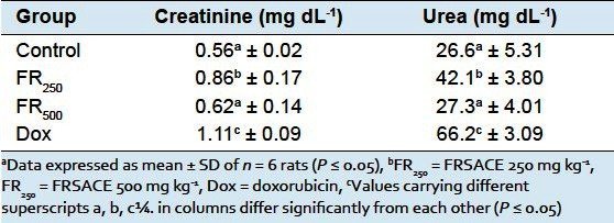
Dox is a known nephrotoxic substance and produces chronic progressive glomerular disease manifested by increased plasma creatinine and urea levels associated with extensive glomerular lesions, tubular dilatation, vacuolization of renal glomeruli, protein deposits in tubular lumen and stromal fibrosis.[4,25] In the present study, serum urea and creatinine levels of control and FR500 pretreated groups were comparable and significantly lower than those of FR250 and Dox groups [Table 1]. Further, no proteinurea was observed in control and FR500 group. Moderate proteinurea was observed in FR250group, while severe proteinurea was found in Dox group. These findings were further supported by the histopathological profiles of the kidneys. The sections from control group showed normal renal tubules associated with normal glomerulus. However, in Dox-treated group focal tubular and glomerular damage leading to shrinkage of the glomerulus was observed. The sections extract pretreated groups showed no tubular damage; however, slight shrinkage of glomerulus was observed [Figure 2]. These observations are in good agreement with earlier reports.[25,29,30].

Table 1 Effect of FRSACE on serum urea and creatinine levels

Figure 2
(a) Section of the kidney of control rats showing normal glomeruli.
(b) Section of the kidney of untreated rats showing focal glomerular and tubular damage with mononuclear infiltrate.
(c) Section of the kidney of FRSACE-treated rats (250 mg kg-1) showing ...

Dox is also known to disturb spermatogenesis leading to low testicular sperm count and associated with increased ROS production.[3] The histopathological section of the control group showed normal seminiferous tubules with good number of sperms, while complete inhibition of spermatogenesis was seen in Dox-treated group, wherein severe damage to the seminiferous tubules with vacuolation and necrosis of the lining epithelial cells and damaged basement membrane resulting in the loss of architecture and spermatogenesis was observed. In FR500 pre-treated group the seminiferous tubules showed normal testicular architecture with sperms. In FR250 pretreated group, although, spermatogenesis was restored toward normalization, the number of sperms was significantly lower compared to FR500 group. These sections also showed vacuolation of the lining epithelium [Figure 3]. The observations are consistent with an earlier report, wherein green tea extract rich in polyphenolic compounds reversed Dox-induced pathological, biochemical changes, histology and lipid peroxidation in rats.[27] The testicular protective activity of FRSACE could be due to the presence of gallic acid and ellagic acid reported to exhibit significant protection against Dox-induced testicular toxicity.[27]

Figure 3
(a) Section of the testis of control rats showing normal seminiferous tubules containing sperms
(b) Section of the testis of untreated rats showing damage to the seminiferous tubules with damaged basement membrane and necrosed lining epithelium,

Oxidative stress was assessed by the levels of glutathione and thiobarbituric acid reactive substances (TBARS) in kidney and testis homogenates. FR250 and FR500 pretreatment significantly decreased (P ≤ 0.05) lipid peroxidation induced by Dox as reflected by lower TBARS and higher GSH values [Figures [Figures44 and and5].5]. It is noteworthy that, FR500 completely reversed oxidative stress to normal levels which could be attributed to the presence of various phenolic compounds and flavonoids such as quercetin, gallic acid, ellagic acid and trepenoidslupeol, lupeol acetate and a-amyrin that are reported to act as strong antioxidant and anti-inflammatory agents.[31]

Figure 4 TBARS levels in kidney and testis. Data expressed as mean ± SD ofn = 6 rats (P ≤ 0.05)

Figure 5 Glutathione levels in kidney and testis. Data expressed as mean ± SD of n = 6 rats (P ≤ 0.05)

Back to Top
Conclusion
From the findings of the present study, it is inferred that doxorubicin exposure results in pronounced oxidative stress, and administration of F. racemosa stem bark extract offers renal and testicular protection through its antioxidant properties. Further, there is a need to identify and isolate the specific bioactive compound(s) from F. racemosa bark for its optimal utilization as a therapeutic agent to derive maximum benefits of doxorubicin as an anticancer drug by reducing its toxic effects considerably.
Back to Top
References

1. Priestman T. Cancer chemotherapy in clinical practice. London: Springer-Verlag; 2008.

2. Ng R, Better N, Green MD. Anticancer agents and cardiotoxicity. Semin Oncol. 2006;33:2–14.[PubMed]

3. Lui RC, Laregina MC, Herbold DR, Johnson FE. Testicular cytotoxicity of intravenous doxorubicin in rats. J Urol. 1986;136:940–3. [PubMed]

4. Injac R, Strukelj B. Recent advances in protection against doxorubicin-induced toxicity. Technol Cancer Res Treat. 2008;7:497–516. [PubMed]

5. Ratain MJ, Ewesuedo RB. Cancer chemotherapy. Berlin: Springer-Verlag; 1999. pp. 76–83.

6. Quiles JL, Huertas JR, Battiono M, Mataix J, Ramírez-Tortosa MC. Antioxidant nutrients and adriamycin toxicity. Toxicology. 2002;180:79–95. [PubMed]

7. Ananymous. The Wealth of India. Vol. 4. New Delhi: Council of Scientific and Industrial Research; 1952. pp. 35–6.

8. Ahmed F, Urooj A. Antioxidant activity of various extracts of Ficus racemosa stem bark. National Journal of Life Sciences. 2009;6:69–74.

9. Ahmed F, Urooj A. Glucose lowering, hepatoprotective and hypolipidemic activity of stem bark ofFicus racemosa in streptozotocin-induced diabetic rats. J Young Pharm. 2009;1:160–4.

10. Ahmed F, Hudeda S, Urooj A. Antihyperglycemic activity of Ficusracemosa bark extract in type II diabetic individuals. J Diabetes. 2010;3:1–2.

11. Ahmed F, Sharanappa P, Urooj A. Antibacterial activities of various sequential extracts of Ficusracemosa stem bark. Pharmacognosy Journal. 2010;2:203–6.

12. Ahmed F, Chandra JN, Manjunath S. Acetylcholine and memory enhancing activity ofFicusracemosa bark. Pharmacognosy Res. 2011;3:246–9. [PMC free article] [PubMed]

13. Ahmed F, Siddesha JM, Urooj A, Vishwanath BS. Radical scavenging and angiotensin converting enzyme inhibitory activities of standardized extracts of Ficusracemosa stem bark. Phytother Res.2010;24:1839–43. [PubMed]

14. Ahmed F, Urooj A. Hepatoprotective effects of Ficusracemosa stem bark against carbon tetrachloride-induced hepatic damage in albino rats. Pharm Biol. 2010;48:210–6. [PubMed]

15. Ahmed F, Urooj A. Cardioprotective activity of standardized extract of Ficusracemosa stem bark against doxorubicin-induced toxicity. Pharm Biol. 2012;50:468–73. [PubMed]

16. Ellman GL. Tissue sulfhydryl groups. Arch Biochem Biophys. 1959;82:70–7. [PubMed]

17. Ohkawa H, Ohishi N, Yagi K. Assay for lipid peroxides in animals tissues by thiobarbituric reaction.Anal Biochem. 1979;95:351–8. [PubMed]

18. Herman EH, Ferrans VJ. Animal models of anthracycline cardiotoxicity: Basic mechanisms and cardioprotective activity. Prog Pediatr Cardiol. 1998;8:49–58.

19. Au WW, Hsu TC. The genotoxic effects of adriamycin in somatic and germinal cells of the mouse.Mutat Res. 1980;79:351–61. [PubMed]

20. Sawada T, Tamada H, Mori J. Secretion of testosterone and epidermal growth factor in mice with oligozoospermia caused by doxorubicin hydrochloride. Andrologia. 1994;26:151–3. [PubMed]

21. Imahie H, Adachi T, Nakagawa Y, Nagasaki T, Yamamura T, Hori M. Effects of adriamycin, an anticancer drug showing testicular toxicity, on fertility in male rats. J Toxicol Sci. 1995;20:183–93.[PubMed]

22. Goodman J, Hochstein P. Generation of free radicals and lipid peroxidation by redox cycling of adriamycin and daunomycin. Biochem Biophys Res Commun. 1977;77:797–803. [PubMed]

23. Pritsos CA, Sokoloff M, Gustafson DL. PZ-51 (Ebselen) in vivo protection against adriamycin-induced mouse cardiac and hepatic lipid peroxidation and toxicity. Biochem Pharmacol. 1992;44:839–41.[PubMed]

24. Shinozawa S, Gomita Y, Araki Y. Protective effects of various drugs on adriamycin (doxorubicin)-induced toxicity and microsomal lipid peroxidation in mice and rats. Biol Pharm Bull. 1993;16:1114–7.[PubMed]

25. Mansour MA, El-Kashef HA, Al-Shabanah OA. Effect of captopril on doxorubicin-induced nephrotoxicity in normal rats. Pharmacol Res. 1999;39:233–7. [PubMed]

26. Manabe F, Takeshima H, Akaza H. Protecting spermatogenesis from damage induced by doxorubicin using the luteinizing hormone-releasing hormone agonist leuprorelin: An image analysis study of a rat experimental model. Cancer. 1997;79:1014–21. [PubMed]

27. Patil LJ, Balaraman R. Green tea extract protects doxorubicin induced testicular damage in rats.Pharmacologyonline. 2008;3:913–25.

28. El-Shenawy NS, Abdel-Nabi IM. Hypoglycemic effect of Cleome droserifolia ethanolic leaf extract in experimental diabetes, and on non-enzymatic antioxidant, glycogen, thyroid hormone and insulin levels. Diabetologia Croatica. 2006;35:15–22.

29. El-Shitany NA, El-Haggar S, El-desoky K. Silymarin prevents adriamycin-induced cardiotoxicity and nephrotoxicity in rats. Food Chem Toxicol. 2008;46:2422–8. [PubMed]

30. Saad SY, Najjar TA, Al-Rikabi AC. The prevention role of deferoxamine against acute doxorubicin-induced cardiac, renal and hepatic toxicity in rats. Pharmacol Res. 2001;43:211–8. [PubMed]

31. Khan N, Sultana S. Chemomodulatory effect of Ficusracemosa extract against chemically induced renal carcinogenesis and oxidative damage response in Wistar rats. Life Sci. 2005;29:1194–210.[PubMed]

Back to Top
Research
 
 
Photo Gallary
Contact us
Address :Rahul Apartment, Satbhai Lane, Dilhi gate, Ahmednagar - 414001
Ph.no.: (0241)2345894
Email: nkortikar@gmail.com